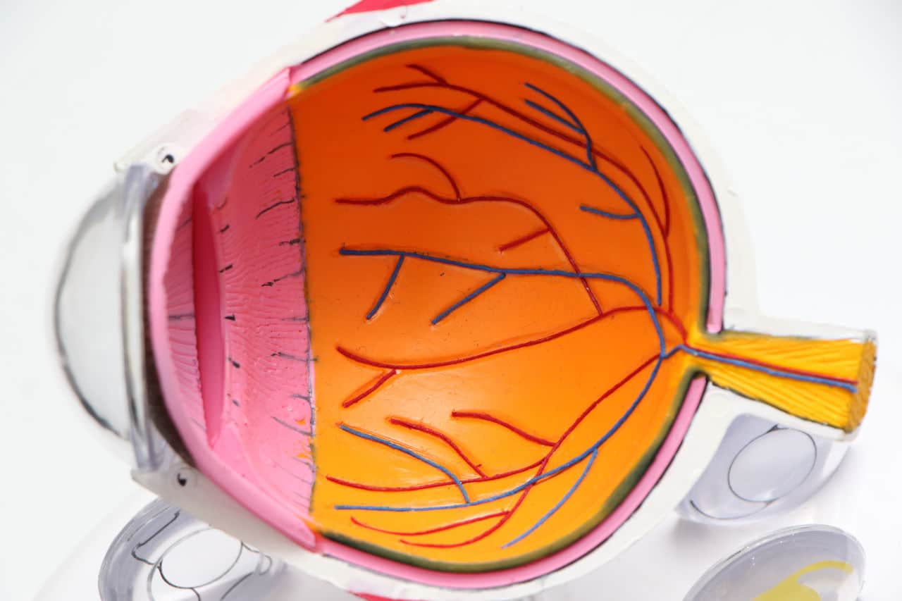
The retina is a thin tissue layer located at the back of the eye. Its duty is to convert images the lens focuses light on to signals. These signals are, in turn, sent to the brain. When there’s retinal detachment, it means that the above cannot take place.
Retinal detachment is said to be a separation of the retina from its usual location; the back of the eye. It is a situation whereby the retina cells are pulled away from the layer of blood vessels, which gives oxygen to the eye and nourishes it. It is a medical emergency and should not be treated with laxity.
If the retina is left removed from its normal position for an extended period, there is a great chance it would result in a permanent loss of sight.
Usually, retinal detachment does not come with pain, so the symptoms are easily overlooked. When an ophthalmologist ignores the signs of this defect, it can lead to grave consequences from delay in treatment. These consequences are not limited to but include the individual having their vision gone permanently.
According to a medical journal by Investigative Ophthalmology and Visual Science (IOVS), retinal detachment (RD) has a modest incidence with a lifetime risk of 3% by age 85. It is classified as a true ophthalmic emergency. This investigation indicates that retinal detachment is a rare condition to a large extent.
However, its rareness does not undermine its severity.
Symptoms of Retinal Detachment
Regarding retinal detachment, the American Academy of Ophthalmology researched the fact that there have been too many misdiagnoses surrounding the defect. This result implies that some symptoms that indicate retinal detachment also indicate other illnesses. It then shows that the ophthalmologists couldn’t note the differences at that point for several reasons.
Retinal detachment can show up as blurry sight and feeling like a covering has been pulled over one’s vision.
The patient may also experience flashes of light that may occur in one or both eyes and a shadowing effect. The most common symptom is seeing tiny bits of debris or black flecks floating before one’s vision (floaters).

Risk Factors of Retinal Detachment
Some factors make an individual liable to have a retinal detachment. Old age is a significant factor, but some of the most prevalent risk factors include having a family history of retinal detachment, especially previous eye surgery, like cataract removal or injury. This information implies that one must be particular about the ophthalmologist who is to carry out the treatment when one has an eye injury.
Also, another reason the medical practitioner must be meticulous is so that they will be able to beat time. Time is of the essence in detecting retinal detachment and likewise in surgeries. If treatment is administered on time, the surgeries have high success rates. Good ophthalmologists know this and strive to get things done on time.
Notably, the primary treatment to reattach a retina is usually surgery. However, the ophthalmologist could use simple procedures in minor detachment cases. Photocoagulation is one of the common treatments for tears in the retina. For photocoagulation, using a laser to burn around the torn area causes a screening and fixes the retina back to its position.
Cryopexy (freezing) is another one.
Here, a freezing probe will be placed in the torn area. This action causes scarring that brings the retina back in place. Other treatment methods include pneumatic retinopexy, vitrectomy, and scleral buckling. These procedures are for the treatment of more prominent forms of retinal detachment.
Case Reference
One of such larger forms of retinal detachment treatment is seen in this case between Nowatske v. Osterloh. The defendant was negligent in treating the plaintiff’s retinal detachment through scleral buckling.
The ophthalmologists are expected to do due diligence by informing the patients of the pros and cons of the surgery they are about to undergo. In this case, the defendant did not notify the plaintiff of the chances of becoming blind through the surgical process beforehand. The defendant also did not use the right equipment while carrying out some procedures during the surgery. Instead, he used his fingers.
After the surgery, the plaintiff couldn’t see in his right eye, and after a post-operative check-up, the defendant concluded that the plaintiff had been made blind permanently in that eye. The ophthalmologist could have easily prevented this complication if the defendant had paid more attention to the treatment procedures.
Likewise, in the case of Stundon v. Stadnik, the defendant performed two cataract extraction surgeries on the plaintiff, after which the plaintiff’s retina got detached. They also permanently lost their vision.
Like the previous case, the defendant caused the plaintiff to lose sight during the cataract operation due to his negligence. He also failed to sufficiently inform the plaintiff of the risks involved in performing eye surgery. The plaintiff’s vision would have been intact if The medical personnel had taken proper care.

Using Legal Animation to Illustrate Errors in Retinal Detachment
The surgical procedures needed to reattach a retina to its rightful position are relatively simple. But because human beings administer them, there are bound to be errors and flaws. Legal animation can help to shed light on errors relating to retinal detachment surgeries.
The use of legal animation cannot be overemphasized when it comes to medical procedures. This claim is because seeing rather than hearing the details of these procedures alone makes comprehension swift and easy. Legal animation would display the anatomy of the human eye and the cause and process by which the retina detached from its usual location.
Let’s take a situation where a previous eye surgery, say, a cataract, causes the retina to be detached as an example. Legal animation would show the presiding judge, jurors, and all present in the court how this previous eye surgery caused the retina to detach from the back of the eye.
They would see the process of detachment and its effect on the individual’s eye. Legal animation will prove how the plaintiff can lose their vision during the surgery. The jury will also see how late detection of the defect poses a significant threat to the individual. They will know how the blood vessels being disconnected from the eyes negatively affects sight.
The surgical procedure would also be displayed, as to how the ophthalmologist went about it and where they went wrong in the treatment process. The court can pick out specific points that could have been avoided or overcompensated.
Conclusion
A retinal detachment animation will undoubtedly be influential in a retinal detachment case. However, the onus is on the attorney on the case to ensure that such animation is created in accordance with the rules of admissibility.
At Fox-AE, having created demonstrative aids for legal use for hundreds of cases, we clearly understand what and what not will make a legal animation admissible from experience. Hence, our medical animators work closely with expert witnesses to ensure that our demonstrative aids reflect the facts and opinions of the case.


