It is medically believed that bones make up about 15 percent of the body of a human being. An average adult has 206 bones in the body in different locations, sizes, strengths, and so on. One of the constant variables common to all the bones in the body, sadly, is that they can all be fractured.
Fractures are complete or partial breaks in bones. It could be an open fracture – where the broken bone is exposed through the skin, as is the case in Hill v. Medlantic HealthCare Group – or closed when the skin is still intact despite the broken bone.
Whether open or closed, fractures are usually caused by twisting or falls from standing height or a higher level of force, like a fall from an extended distance or even a car accident, or anytime the bone encounters more weight than it is able to bear.
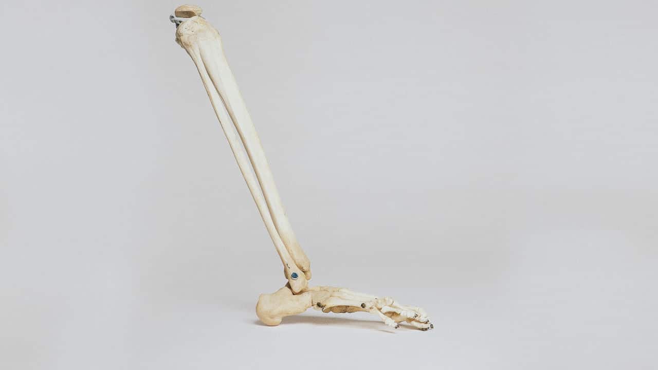
The tibia (also called the shin bone) is one of the two long bones found in the lower part of the leg alongside the fibula. The tibia, however, is the larger bone located on the inside and bears more weight than the fibula, which is smaller, found on the outside, and bears less weight.
The National Center for Health Statistics reports 492,000 tibial fractures per year in the United States.
Causes of Tibial Fracture.
Of the hundreds of thousands of tibial fracture cases recorded annually, the causes can either be dramatic events or some not-so-spectacular circumstances.
The latter is the case in Moore v Singh where a little girl earlier complained of consistent pain in her leg. The first attempt to diagnose what went wrong did not show the presence of a tibial fracture, and a rather simple treatment was administered. It was not until some weeks later that she was diagnosed with a tibial fracture on the same leg.
However, tibia fractures, in most cases, are caused by traumatic injuries, like motor vehicle accidents or falls.
In Reynolds v Gonzalez, the plaintiff, a twenty-seven years old man, sustained serious injuries to his left leg and had his tibia fractured in a dirt-bike accident.
In addition, tibial fractures can happen during sports activities that require contact, especially those that involve repeated involvement of the shin bones, such as long-distance running, kickboxing, American football, etc.
An example is John Herlich v. Jason Srouy, where the plaintiff, a referee of American football, claimed to have been injured by one of the players, and he sustained a tibial and fibula fracture.
Osteoporosis, a medical condition that makes the bones weaker than usual, is another common cause of tibial fracture.
Diagnosing Tibial Fracture
A 3D animation can be used to give an interactive model of the processes involved in diagnosing tibial fracture in a case involving such circumstances. This mostly employs a physical examination.
More sophisticated and medically advanced test mechanisms would be the X-ray, which involves the use of unseen electromagnetic energy beams to make images of the bones to diagnose infections and bone injuries in a person’s leg or hands.
Magnetic Resonance Imaging (MRI) is also another imaging test that employs the use of big magnets, a computer, and radiofrequency to produce a detailed image of the structures in the body.
In addition, Computed Tomography (Ct) Scan, which is more effective than an X-ray and gives a 3-D image of the bone, can also be used.
Using Legal Animation In A Tibial Fracture Case
In Johnson v Jamaica Hospital Medical Center, the plaintiff commenced an action to recover damages for medical malpractice and other charge counts.
After his gunshot wound was treated with fixation, he was discharged, having spent just four days in the hospital. However, two months later, he developed a serious infection in the leg that made him stay 63 days, and had to go through seven different surgeries.
In addition, the case of Reynolds v MAINEGENERAL HEALTH would not have been subjected to a review if the counsel of the plaintiff-appellant had employed a visual animation in proving the medical malpractice of the defendant-respondent. The plaintiff was involved in a ghastly accident, after which he was immediately rushed to the defendant’s hospital.
Upon appeal, the US Court of Appeals affirmed the summary judgment of the district court discounting the plaintiff’s claim of misdiagnoses and negligent treatment from the medical practitioners of the defendant’s hospital.
Conclusion
Whether for mediation or an outright trial of a case involving a tibial fracture, to have an easily understandable inkling of the injury, the possible causes, the expected treatment to be applied, and to establish whether or not there is medical malpractice or negligence, a counsel can employ the use of tibial fracture animation to illustrate the facts of the case.
The expert opinions and the medical jargon involved will be illustrated in the simplest of terms – as a 3D animation of the tibial fracture will provide a graphic evaluation and management of tibial fractures and illustrate the role of interprofessional team members in working together to provide well-coordinated care and enhance victim outcomes.
However, such an animation must be created in accordance with the rules of admissibility for demonstrative evidence.


